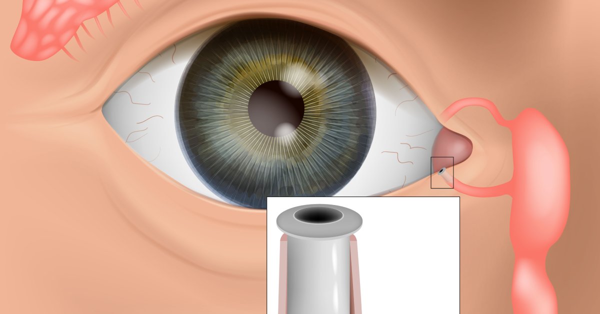
Punctal plugs are a treatment option for managing dry eye syndrome. They function by blocking the tear ducts (puncta) to conserve tears, providing relief for patients with insufficient natural tear production. It’s required for medical professionals to know how to insert and remove punctal plugs safely. For optimal patient outcomes while minimizing risks, here are the steps involved in the procedure.
Prepare for the Procedure
Preparation is necessary for a safe and effective procedure. The healthcare professional assesses the patient’s suitability for punctal plugs. They also consider the patient’s medical history to identify any contraindications, such as infections, punctal abnormalities, or allergies to the plug materials. There must be assurance the chosen plug type is appropriate based on the patient’s condition, whether it’s temporary (collagen) or permanent (silicone).
A sterile environment is necessary to reduce the risk of infection. Preparing the required equipment, including sterile forceps, magnification tools, and punctual plugs, is a part of the process. Anesthetic eye drops should also be on hand to minimize the patient’s discomfort.
Safe Plug Insertion
Eye care professionals can use topical anesthetic drops to alleviate discomfort. Magnification tools help with visualizing the punctal area clearly. In the case of a constricted punctum, gentle dilation may be necessary before placing the plug.
Using sterile forceps, align the plug with the punctum and gently insert it. The plug should rest slightly below the punctual rim to prevent extrusion without causing canalicular damage.
Once in place, verify its position to ensure it’s secure and comfortable for the patient. Post-procedure consulting is important, so the patient is informed on potential sensations of foreign body awareness and any signs of complications to monitor, such as redness or persistent discomfort.
Plug Removal Safety
For medical professionals, the primary focus during punctal occlusion is how to insert and remove punctal plugs safely. Before proceeding, the eye care professional will confirm the necessity of removal and outline the process to the patient. Removing punctual plugs may occur due to over-retention of tears, discomfort, or a need to switch to a different treatment option.
The process begins with topical anesthetic drops. If the plug is visible, sterile forceps gently grip the exposed portion. Avoid excessive force or twisting; these actions could damage the punctual tissues.
For plugs that have migrated deeper, more advanced techniques, such as irrigation, may be required. Exercise caution to avoid trauma during this process.
After removal, evaluate the punctal area for irritation or damage. Patients will receive post-procedure instructions. Advisement usually includes reporting any discomfort or unusual symptoms immediately.
Patient Safety and Care
The success of punctal plug procedures hinges on meticulous attention to detail. The focus on sterility helps prevent infections, as well as the vigilance of eye care professionals in checking for signs of complications. Effective communication helps patients feel reassured and increases cooperation during these procedures.
Investing in high-quality, punctual plugs for sale from suppliers, such as Automated Ophthalmics, that manufacture in-house minimizes the risk of quality control issues. By adhering to these guidelines, eye care professionals are able to provide a safe and comfortable experience while achieving optimal therapeutic outcomes.